|
|
|
|
Atlanta Breast Wellness
Thermography Atlanta
Atlanta Body Care
(770) 355-8352
Call to Schedule an appointment
|
Introducing the First
Pain-free, Radiation-free
Digital Breast Exam

|
|
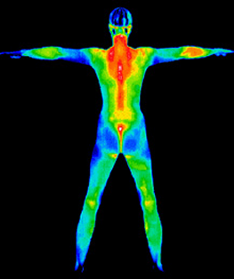
Digital Infared Thermal Imaging-Thermography
SureTouch Breast Exam
Ultrasound
Shoe Orthotics

Check, HSA Card, Debit Card, or Zelle
All cards (3% service fee)
(if you require other forms of payment please let me know)
|
ANNOUNCEMENT
Good News
I Have moved back to Atlanta.
As of September 1, 2021
I will be providing services in Atlanta/Roswell.
500 Sun Valley Dr. Suite D-1 Roswell, GA. 30076
It has been my pleasure serving the Atlanta community for the past 20 yrs, And. I am please to announce my return on a full time basis to provide services in the Atlanta area.
Please reach out to me when you are ready to schedule your appointment.
If you have any questions, please contact me by phone, text or email.
BLESSINGS and Thank You for your loyalty.
Dr. Rosalind Gamba, NMD,
Phone: c- 770-355-8352 email: roz158@bellsouth.net
|
|
|
 |
 |
Breast Questions & Answers
|
|
|
Digital Infrared Thermal Imaging (DITI) offers the opportunity of earlier detection of breast disease than has been possible with breast self-examination, physician palpation or mammography alone.
Each individual has her own thermal pattern (normally symmetric) that is accurate and static throughout her lifetime. Any changes to her normal “thermal fingerprint” caused by early cell changes (pathology) will become increasingly apparent. Monitoring changes over periods of time with DITI is the most efficient means of identifying subjects who require further investigation.
DITI is a non-invasive test. There is no contact with the body of any kind, no radiation and the procedure is painless. The scanning system merely detects and records the infrared radiation that is emitting from the patient’s body.
Utilizing sophisticated infrared technology and innovative computer software, thermal imaging technicians simply capture a digitized image of the breast in the form of an infrared thermogram, or heat picture. |
|
|
|
Canadian researchers recently confirmed that infrared imaging of breast cancers could detect minute temperature variations related to blood flow and demonstrate abnormal patterns associated with the progression of tumors. These images, or thermograms of the breast, were positive for 83% of breast cancers compared to 61% for clinical breast examination alone and 84% for mammography. The 84% sensitivity rate of mammography alone was increased to 95% when infrared imaging was added.
|
|
|
|
Yes. Unlike mammography and ultrasound, Digital Infrared Thermal Imaging (DITI) is a test of physiology. It detects and records the infrared heat radiating from the surface of the body. It can help in early detection and monitoring of abnormal physiology and the establishment of risk factors for the development or existence of cancer.
Mammography and ultrasound are tests of anatomy. They look at structure. When a tumor has grown to a size that is large enough and dense enough to block an x-ray beam (mammography) or sound wave (ultrasound), it produces an image that can be detected by a trained radiologist.
Neither mammogram, ultrasound, nor DITI can diagnose cancer. Only a biopsy can diagnose cancer. But, when DITI, mammograms, ultrasounds, and clinical exams are used together, the best possible evaluation of breast health can be made. |
|
|
|
No. While some women make a personal choice to use thermal imaging instead of mammography for breast screening, other women who cannot use mammography for a number of reasons can use thermography instead of mammography. Most women use thermal imaging in addition to mammography and/or ultrasound.
We believe that (DITI) should be viewed as a complementary, not competitive, tool to mammography and ultrasound. DITI has the ability to identify patients at the highest level of risk and actually increase the effective usage of mammograms and ultrasounds. Research confirms that DITI, when used with mammography, can improve the sensitivity of breast cancer detection.
The ultimate choice should be made on an individual basis with regard to clinical history, personal circumstances and medical advice. |
|
|
|
No. DITI detects and records the infrared heat radiating from the surface of the body. There is no contact with the body or harmful radiation. |
|
|
|
No. There is no contact with the body or painful breast compression. |
|
|
|
Any adult can have a thermal breast scan. This test is designed to improve chances for detecting fast growing tumors in the intervals between mammographic screenings or when mammography is not indicated by screening guidelines for women under 50.
DITI is especially appropriate for younger women under 50 years whose denser breast tissue makes it more difficult for mammography to pick up suspicious lesions. This test can provide a ‘clinical marker’ to the doctor or mammographer, indicating that a specific area of the breast needs closer examination.
Breast cancers tend to grow significantly faster in younger women (under 50 years). The average tumor doubling time for women under 50 is 80 days compared to 157 days for women between 50 – 70 years. Secondly, the faster a malignant tumor grows, the more infrared radiation it generates. Therefore, for younger women in particular, results from DITI screening can lead to earlier detection. |
|
|
|
Yes. The information provided by a thermography study can contribute useful additional information which ultimately helps your doctor with case management decisions. It is also important to establish a baseline for future comparison in order to monitor changes and the progress of any treatment. |
|
|
|
Once a reliable baseline has been established, which normally requires two studies 3-months apart, you should have an on-going annual comparative study to detect any suspicious functional (physiological) changes, warranting further investigation. Depending on your personal history and risk for breast disease, your doctor can advise how often you should have a thermal scan repeated. |
|
|
|
Yes! Over 800 peer-reviewed studies on breast thermography exist in the index medicus literature. In this database, well over 300,000 women have been included as study participants. The numbers of participants in many studies are very large (10,000, 37,000, 60,000, 85,000, etc.) Some of these studies have followed patients for up to 12 years.
These clinical trials have demonstrated that breast thermography:
- detects the first signs of a cancer up to 10 years before any other procedure can detect it
- significantly augments the long-term survival rates of its recipients by as much as 61%
- when used as part of a multimodal approach (clinical examination + mammography + thermography), will detect 95% of early stage cancers
|
|
|
|
When thermography was first explored for breast imaging, it was viewed as competitive to mammograms. It was tested and evaluated to see if it was safer and more diagnostically accurate than mammography. These comparisons should not have been made, as you can not compare tests of physiology and anatomy.
In particular, when thermography was tested on younger women, thermographic abnormalities were detected many times but mammograms did not detect any tumors. The results were considered “false positives”. The more patients of younger age screened with the so-called false positive, the more suspicion was placed on thermography. Years later, in re-call studies, a large percentage of these women had developed breast cancer or other breast disease, in the exact location of the abnormal “false-positive” thermogram, thus validating its early warning role. Thermography’s only “error” was that it was too accurate too early and the results couldn’t be corroborated at the time.
Secondly, thermography was being used in sports medicine, dentisty, podiatry, chiropractic, orthopedics rheumatology, and neurology in a variety of support or adjunctive diagnostic roles. It was soon realized that thermography could clearly, objectively, and easily demonstrate the physiological component of pain and injury, especially to the spinal column, due to car accidents, job injuries, and a host of other “tort” related law suits. Everyone involved had benefited from these positive test findings, which could be clearly shown to a jury. Everyone that is except the defendant insurance industry.
Needless to say, the insurance industry in the United States placed an all-out effort to diminish the value of thermography in courts of law due to high litigation costs. Eventually, lobbying efforts at the AMA’s House of Delegates and at Medicare, brought about the removal of thermographic coverage by most insurance companies and the greatly reduced utilization of thermography in the United States. This was most unfortunate for the patients who could clearly benefit from thermal imaging. |
|
|
 |
|
 |
|
|